
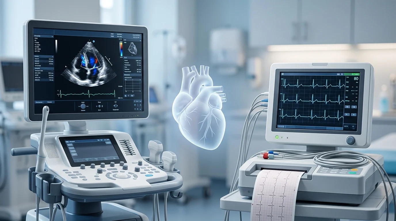
Quick Answer: An echocardiogram uses ultrasound waves to create images of the heart, while an EKG (electrocardiogram) records the heart’s electrical activity. An echocardiogram helps doctors evaluate heart structure, valves, and pumping function. In contrast, an EKG helps detect abnormal heart rhythms and electrical problems. Doctors may use both tests to assess heart health.
Heart tests help doctors understand how well your heart works and identify possible problems. Two common tests are an echocardiogram and an EKG. Although both evaluate heart health, they examine different aspects of the heart.
An echocardiogram produces moving images of the heart using sound waves. It allows doctors to examine the heart’s chambers, valves, and pumping ability. An EKG, on the other hand, records electrical signals that control each heartbeat. This test can help identify irregular rhythms and other electrical abnormalities.
So, which test do you need? The answer depends on your symptoms and your doctor’s concerns. For example, a doctor may order an EKG for an irregular heartbeat but recommend an echocardiogram to investigate a heart murmur or suspected valve problem.
Understanding these differences can help you know what to expect and why your doctor may recommend one or both tests.
What Is an Echocardiogram?
An echocardiogram, often called an echo, is an imaging test that uses high-frequency sound waves to create pictures of your heart.
During a standard echocardiogram, a technician applies gel to your chest and moves a small device called a transducer over your skin. The device sends sound waves into your body and captures their echoes to create heart images.
Doctors use these images to assess:
- Heart size and shape
- Heart valve movement and function
- How effectively the heart pumps blood
- Blood flow through the heart
- Certain structural abnormalities
A standard transthoracic echocardiogram is usually painless and takes around 30 to 60 minutes. It does not use radiation.
What Is an EKG?
An EKG, also called an ECG, stands for electrocardiogram. It records the electrical signals that make your heart beat.
During the test, a healthcare professional attaches small adhesive electrodes to your chest and sometimes your arms and legs. These electrodes detect electrical activity and send the information to a machine, which displays the results as a series of lines on a graph.
An EKG can help detect:
- Irregular heart rhythms, also called arrhythmias
- A fast or slow heart rate
- Certain signs of a heart attack
- Electrical conduction problems
- Some changes that may suggest heart muscle strain
A standard resting EKG usually takes about 5 to 10 minutes, although preparation may take additional time. The test is painless and does not send electricity into your body.
Echocardiogram vs EKG: Key Differences
| Feature | Echocardiogram | EKG |
|---|---|---|
| Main purpose | Examines heart structure and function | Records electrical activity |
| Technology | Ultrasound waves | Electrodes and electrical signal recording |
| Shows heart images | Yes | No |
| Detects irregular rhythms | Not its main purpose | Yes |
| Evaluates heart valves | Yes | Not directly |
| Measures pumping function | Yes | Not directly |
| Typical test time | 30 to 60 minutes | 5 to 10 minutes |
| Radiation | No | No |
Although these tests provide different information, doctors may use them together to build a clearer picture of your heart health.
When Do You Need an Echocardiogram or an EKG?
Your doctor chooses a test based on your symptoms, medical history, and physical examination.
An echocardiogram may be recommended when:
- Your doctor hears an unusual heart murmur.
- You have symptoms that may suggest heart failure.
- A heart valve problem is suspected.
- Your doctor needs to assess heart pumping function.
- You have a known structural heart condition that needs evaluation.
An EKG may be recommended when:
- You experience palpitations or a racing heartbeat.
- You feel dizzy or faint.
- Your doctor suspects an abnormal heart rhythm.
- You have chest discomfort that needs assessment.
- Your healthcare team needs to evaluate your heart’s electrical activity.
However, neither test identifies every heart condition. For example, a normal EKG does not rule out every heart problem, and an echocardiogram may not detect intermittent rhythm abnormalities. Therefore, your doctor may recommend additional testing.
Can You Have Both Tests?
Yes. Doctors often order both tests because they provide complementary information.
For instance, if you experience shortness of breath and palpitations, an EKG may help identify an electrical problem. Meanwhile, an echocardiogram can show whether your heart’s structure or pumping function is abnormal.
Depending on your symptoms, your doctor may also recommend blood tests, a stress test, or a Holter monitor, which records heart rhythms over a longer period.
Common Misconceptions
1. An echocardiogram and an EKG are the same test.
They are different. An echocardiogram creates heart images, whereas an EKG records electrical signals.
2. A normal EKG means your heart is completely healthy.
Not necessarily. Some structural problems and intermittent heart conditions may not appear on a resting EKG.
3. An echocardiogram can detect every heart problem.
No. Although it provides valuable structural information, it cannot detect every electrical, coronary artery, or other heart condition.
Frequently Asked Questions
Is an echocardiogram more accurate than an EKG?
Neither test is universally more accurate. Each evaluates different aspects of heart health, so the appropriate test depends on the suspected condition.
Which test is better for detecting blocked arteries?
Neither test directly shows all coronary artery blockages. Doctors may recommend other tests, such as coronary CT angiography or a stress test, depending on the situation.
Does an EKG show heart damage?
An EKG can reveal changes that suggest a heart attack or other heart muscle problems. However, doctors may need blood tests and imaging to confirm the cause.
Is an echocardiogram painful?
A standard echocardiogram is generally painless. You may feel slight pressure from the transducer against your chest.
Do I need to prepare for either test?
A standard resting EKG usually requires little preparation. A routine echocardiogram also typically requires no special preparation, although instructions can vary by test type.
Can an EKG detect heart failure?
An EKG may show abnormalities associated with heart failure, but it cannot confirm or rule out the condition by itself. An echocardiogram often helps assess heart pumping function.
How long do the results take?
A healthcare professional can often review an EKG shortly after the test. Echocardiogram images may require interpretation by a qualified clinician, so the timing of results varies.
Which test should I ask my doctor for?
Describe your symptoms and let your doctor determine which test is appropriate. The right choice depends on your medical history and the condition being investigated.
Conclusion
An echocardiogram and an EKG serve different but complementary purposes. An echocardiogram shows the heart’s structure, valves, and pumping function, while an EKG records its electrical activity and helps detect rhythm problems.
Ultimately, your doctor may recommend one test or both, depending on your symptoms. If you experience new or severe chest pain, difficulty breathing, or fainting, seek urgent medical care rather than waiting for a routine test.